
Cefalometrijske promjene povezane s MAD terapijom
DOI:
https://doi.org/10.55378/rv.47.2.1Ključne riječi:
opstruktivna apneja u snu, uređaj za pomicanje mandibule, lateralna cefalometrija, nuspojave, gornji dišni putoviSažetak
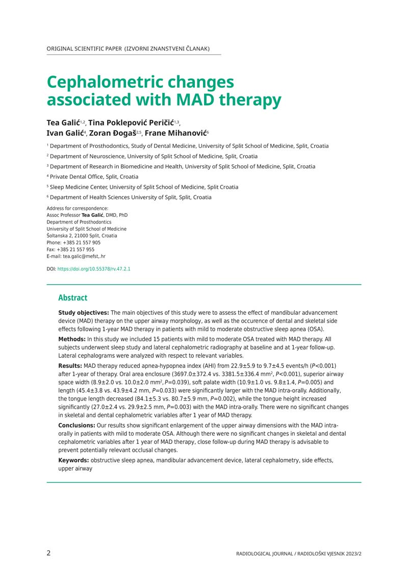
Ciljevi istraživanja: Glavni ciljevi ovog istraživanja bili su procijeniti učinak terapije uređajem za pomicanje mandibule (MAD) na morfologiju gornjih dišnih putova, kao i pojavu dentalnih i skeletnih nuspojava nakon jednogodišnje MAD terapije u bolesnika s blagom do umjerenom opstruktivnom apnejom u snu (OSA).
Metode: U ovo istraživanje uključili smo 15 bolesnika s blagom do umjerenom OSA liječenih MAD terapijom. Svi ispitanici podvrgnuti su studiji spavanja i lateralnoj cefalometrijskoj radiografiji na početku i nakon godinu dana praćenja. Lateralni cefalogrami analizirani su s obzirom na relevantne varijable.
Rezultati: MAD terapija smanjila je indeks apneje-hipopneje (AHI) s 22,9±5,9 na 9,7±4,5 događaja/h (P<0,001) nakon jednogodišnje terapije. Zatvorenost oralnog područja (3697,0±372,4 prema 3381,5±336,4 mm2, P<0,001), širina gornjeg dišnog prostora (8,9±2,0 prema 10,0±2,0 mm2, P=0,039), širina mekog nepca (10,9±1,0 prema 9,8±1,4, P=0,005) i duljina (45,4±3,8 prema 43,9±4,2 mm, P=0,033) bili su značajno veći kod MAD intraoralne primjene. Osim toga, duljina jezika se smanjila (84,1±5,3 prema 80,7±5,9 mm, P=0,002), dok se visina jezika značajno povećala (27,0±2,4 prema 29,9±2,5 mm, P=0,003) kod MAD intraoralne primjene. Nije bilo značajnih promjena u skeletnim i dentalnim cefalometrijskim varijablama nakon 1 godine MAD terapije.
Zaključci: Naši rezultati pokazuju značajno povećanje dimenzija gornjih dišnih putova s MAD intraoralno kod pacijenata s blagim do umjerenim OSA. Iako nije bilo značajnih promjena u skeletnim i dentalnim cefalometrijskim varijablama nakon 1 godine MAD terapije, preporučuje se pomno praćenje tijekom MAD terapije kako bi se spriječile potencijalno relevantne okluzalne promjene.
Reference
Epstein LJ, Kristo D, Strollo PJ Jr, et al. Clinical guideline for the evaluation, management and long-term care of obstructive sleep apnea in adults. J Clin Sleep Med. 2009;5:263-276.
Bassetti C, Dogas Z, Peigneux P, eds. Sleep Medicine Textbook. Regensburg: European Sleep Research Society; 2014.
Jordan AS, McSharry DG, Malhorta A. Adult obstructive sleep apnoea. Lancet. 2014;383:736-747.
Phillips CL, Grunstein RR, Darendeliler MA, et al. Health outcomes of continuous positive airway pressure versus oral appliance treatment for obstructive sleep apnea. Am J Resp Crit Care Med. 2013;187:879-887.
Bozic J, Galic T, Supe-Domic D, et al. Morning cortisol levels and glucose metabolism parameters in moderate and severe obstructive sleep apnea patients. Endocrine. 2016;53:730-739.
Quan SF, Chan CS, Dement WC, et al. The association between obstructive sleep apnea and neurocognitive performance-the Apnea Positive Pressure Long-term Efficacy Study (APPLES). Sleep. 2011;34:303-314.
Bucks RS, Olaithe M, Eastwood P. Neurocognitive function in obstructive sleep apnoea: A meta-review. Respirology. 2013;18:61-70.
Kushida CA, Nichols DA, Holmes TH, et al. Effects of continuous positive airway pressure on neurocognitive function in obstructive sleep apnea patients: the Apnea Positive Pressure Long-term Efficacy Study (APPLES). Sleep. 2012;35:1593-1602.
Sutherland K, Vanderveken OM, Tsuda H, et al. Oral appliance treatment for obstructive sleep apnea: an update. J Clin Sleep Med. 2014;10:215-227.
Ramar K, Dort LC, Katz SG, et al. Clinical Practice Guideline for the Treatment of Obstructive Sleep Apnea and Snoring with Oral Appliance Therapy: An Update for 2015. J Clin Sleep Med. 2015;11:773-827.
Marklund M. Update on oral appliance therapy for OSA. Curr Sleep Med Rep.2017;3:143-151.
Marklund M. Long-term efficacy of an oral appliance in early treated patients with obstructive sleep apnea. Sleep Breath. 2016;20:689-694.
Sawyer AM, Gooneratne NS, Marcus CL, Ofer D, Richards KC, Weaver TE. A systematic review of CPAP adherence across age groups: clinical and empiric insights for developing CPAP adherence interventions. Sleep Med Rev.2011;15:343-356.
Bozic J, Galic T, Supe-Domic D, et al. Morning cortisol levels and glucose metabolism parameters in moderate and severe obstructive sleep apnea patients. Endocrine. 2016;53:730-739.
Anandam A, Patil M, Akinnusi M, Jaoude P, El-Solh AA. Cardiovascular mortality in obstructive sleep apnoea treated with continuous positive airway pressure or oral appliance: an observational study. Respirology. 2013;18:1184-1190.
Naismith SL, Winter VR, Hickie IB, Cistulli PA. Effect of oral appliance therapy on neurobehavioral functioning in obstructive sleep apnea: a randomized controlled trial. J Clin Sleep Med. 2005;1:374-80.
Galic T, Bozic J, Pecotic R, Ivkovic N, Valic M, Dogas Z. Improvement of Cognitive and Psychomotor Performance in Patients with Mild to Moderate Obstructive Sleep Apnea Treated with Mandibular Advancement Device: A Prospective 1-Year Study. J Clin Sleep Med. 2016;12:177-186.
Poirrier AL, Fanielle J, Bruwier A, Chakar B, Poirrier R. Upper airway imaging in sleep-disordered breathing. Acta Neurol Belg. 2014;114:87-93.
Denolf PL, Vanderveken OM, Marklund ME, Braem MJ. The status of cephalometry in the prediction of non-CPAP treatment outcome in obstructive sleep apnea patients. Sleep Med Rev. 2016;27:56-73.
Muretić Ž, Lauc T, Ferreri S. Rendgenska kefalometrija. Zagreb: Školska knjiga; 2014. [In Croatian].
Sutherland K, Deane SA, Chan AS, et al. Comparative effects of two oral appliances on upper airway structure in obstructive sleep apnea. Sleep. 2011;34:469-477.
Sutherland K, Chan AS, Cistulli PA. Three-dimensional assessment of anatomical balance and oral appliance treatment outcome in obstructive sleep apnoea. Sleep Breath. 2016;20:903-910.
Chan AS, Sutherland K, Schwab RJ, et al. The effect of mandibular advancement on upper airway structure in obstructive sleep apnoea. Thorax. 2010;65:726-732.
Pae EK, Ferguson KA. Cephalometric characteristics of nonobese patients with severe OSA. Angle Orthod. 1999;69:408-412.
Mostafiz W, Dalci O, Sutherland K, et al. Influence of oral and craniofacial dimensions on mandibular advancement splint treatment outcome in patients with obstructive sleep apnea. Chest. 2011;139:1331-1339.
Tsuiki S, Isono S, Ishikawa T, Yamashiro Y, Tatsumi K, Nishino T. Anatomical balance of the upper airway and obstructive sleep apnea. Anesthesiology. 2008;108:1009-1015.
Sforza E, Bacon W, Weiss T, Thibault A, Petiau C, Krieger J. Upper airway collapsibility and cephalometric variables in patients with obstructive sleep apnea. Am J Respir Crit Care Med. 2000;161:347-352.
Genta PR, Schorr F, Eckert DJ, et al. Upper airway collapsibility is associated with obesity and hyoid position. Sleep. 2014;37:1673-1678.
Vidović N, Mestrović S, Dogas Z, et al. Craniofacial morphology of Croatian patients with obstructive sleep apnea. Coll Antropol. 2013;37:271-279.
Fritsch KM, Iseli A, Russi EW, Bloch KE. Side effects of mandibular advancement devices for sleep apnea treatment. Am J Respir Crit Care Med. 2001;164:813-818.
Martínez-Gomis J, Willaert E, Nogues L, Pascual M, Somoza M, Monasterio C. Five years of sleep apnea treatment with a mandibular advancement device. Side effects and technical complications. Angle Orthod. 2010;80:30-36.
Almeida FR, Lowe AA, Sung JO, Tsuiki S, Otsuka R. Long-term sequellae of oral appliance therapy in obstructive sleep apnea patients: Part 1. Cephalometric analysis. Am J Orthod Dentofacial Orthop. 2006;129:195-204.
Doff MH, Veldhuis SK, Hoekema A, et al. Long-term oral appliance therapy in obstructive sleep apnea syndrome: a controlled study on temporomandibular side effects. Clin Oral Investig. 2012;16:689-697.
Ringqvist M, Walker-Engström ML, Tegelberg A, Ringqvist I. Dental and skeletal changes after 4 years of obstructive sleep apnea treatment with a mandibular advancement device: a prospective, randomized study. Am J Orthod Dentofacial Orthop. 2003;124:53-60.
Pliska BT, Nam H, Chen H, Lowe AA, Almeida FR. Obstructive sleep apnea and mandibular advancement splints: occlusal effects and progression of changes associated with a decade of treatment. J Clin Sleep Med. 2014;10:1285-1291.
Doff MH, Finnema KJ, Hoekema A, Wijkstra PJ, de Bont LG, Stegenga B. Long-term oral appliance therapy in obstructive sleep apnea syndrome: a controlled study on dental side effects. Clin Oral Investig. 2013;17:475-482.
Doff MH, Hoekema A, Pruim GJ, Huddleston Slater JJ, Stegenga B. Long-term oral-appliance therapy in obstructive sleep apnea: a cephalometric study of craniofacial changes. J Dent. 2010;38:1010-1018.
Almeida FR, Lowe AA, Sung JO, Tsuiki S, Otsuka R. Long-term sequellae of oral appliance therapy in obstructive sleep apnea patients: Part 1. Cephalometric analysis. Am J Orthod Dentofacial Orthop. 2006;129:195-204.
Pecotic R, Dodig IP, Valic M, Ivkovic N, Dogas, Z. The evaluation of the Croatian version of the Epworth sleepiness scale and STOP questionnaire as screening tools for obstructive sleep apnea syndrome. Sleep Breath. 2012;16:793-802.
Tsuiki S, Kobayashi M, Namba K, et al. Optimal positive airway pressure predicts oral appliance response to aleep apnoea. Eur Respir J. 2010;35:1098-1105.
Vanderveken OM, Dieltjens M, Wouters K, De Backer W, Van de Heyning PH, Braem MJ. Objective measurement of compliance during oral appliance therapy for sleep-disordered breathing. Thorax. 2013;68:91-96.
Tsuiki S, Hiyama S, Ono T, et al. Effects of a titratable oral appliance on supine airway size in awake non-apneic individuals. Sleep. 2001;24:554-560.
Hiyama S, Ono T, Ishiwata Y, Kuroda T. Supine cephalometric study on sleep-related changes in upper-airway structures in normal subjects. Sleep. 2000;23:783-790.
Hiyama S, Tsuiki S, Ono T, Kuroda T, Ohyama K. Effects of mandibular advancement on supine airway size in normal subjects during sleep. Sleep. 2003;26:440-445.
Ryan CF, Love LL, Peat D, Fleetham JA, Lowe AA. Mandibular advancement oral appliance therapy for obstructive sleep apnoea: effect on awakecalibre of the velopharynx. Thorax. 1999;54:972-977.
Geoghegan F, Ahrens A, McGrath C, Hägg U. An evaluation of two different mandibular advancement devices on craniofacial characteristics and upper airway dimensions of Chinese adult obstructive sleep apnea patients. Angle Orthod. 2015;85:962-968.
Ghazal A, Jonas IE, Rose EC. Dental side effects of mandibular advancement appliances – a 2-year follow-up. J Orofac Orthop. 2008;69:437-447.
Hamoda MM, Kohzuka Y, Almeida FR. Oral appliances for the management of OSA: An updated review of the literature. Chest. 2017; doi: 10.1016/j.chest.2017.06.005. [Epub ahead of print].
Preuzimanja
Objavljeno
Broj časopisa
Rubrika
Kategorije
Licenca
Autorska prava (c) 2023 Tea Galić, Tina Poklepović Peričić, Ivan Galić, Zoran Dogas, Frane Mihanović (Autor)

This work is licensed under a KreativniCommons Attribution-NonCommercial Međunarodne licence.
 HDRT